Pilonidal Cyst: Etiology, Surgical Interventions and Management
Edited by SarMal, Jen Moreau, Sharingknowledge
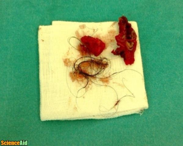
A pilonidal cyst or pilonidal sinus is an abscess (sinus, small hole or tunnel) that develops along the tailbone (coccyx) near the cleft of the buttocks. A pilonidal cyst is generally located 5cm-6cm above the anus. A pilonidal cyst has a distinguishing epithelial track (the sinus) located in the skin of the natal cleft, a short distance behind the anus. This sinus track generally contains hair; as such the term pilonidal is derived from the Latin words "pilus" (hair) and "nidus" (nest). Pilonidal sinuses were first described in 1880 by Hodges, and was a common condition in drivers during World War II. During World War II, 80,000 U.S. soldiers were diagnosed with pilonidal cysts. Researchers believed the condition was caused by riding in bumpy Jeeps and so became known as Jeep Disease. Additional research has demonstrated appropriate management and some methods of preventing pilonidal cysts, but currently, the cause of pilonidal cysts remains unknown.
Incidence
Pilonidal cysts generally occur after puberty and before the age of 40. Pilonidal cysts generally affect males more frequently than females, this is believed to be due to the more hirsute nature of men. [1]. Rick factors associated with pilonidal cysts include:
- Men. Recent research and studies have demonstrated men are almost 10 times more likely that females to develop a pilonidal cyst.
- Caucasians. It is believed that this is due to differing hair characteristics and growth patterns.
- Sedentary Lifestyle or Occupation. Truck drivers, cab drivers and people with sedentary lifestyles develop pilonidal cysts more frequently.
- Obesity. Individuals who are obese, particularly men, are more at risk to the development of pilonidal cysts.
- Family History. Researchers are not exactly sure the connection between family history and the development of pilonidal cysts, but there are theories that because hair growth location, patterns, and characteristics are inherited, an increased risk for pilonidal cyst development occurs.
- Previous Pilonidal Sinus. Patients who have already had a pilonidal cyst are at increased risk for the recurrence of another pilonidal cyst. A 2007 study demonstrated that "pilonidal cyst disease recurred in 41 of 205 patients (20%)," [2].
What Causes a Pilonidal Cyst?
The cause of pilonidal cysts remains unknown, however many theories exist. Many of these theories were developed after World War II in response to the 80,000 U.S. soldiers diagnosed with the condition. Following the war, Patey and Scarf hypothesized pilonidal cysts were instigated by the presence of hair into subcutaneous tissue. The hair forces itself into the subcutaneous tissue and in doing so creates a granuloma, increased granulation tissue or a swelling of the surrounding cells.
More recent research asserts pilonidal cysts are caused in the epidermis when disrupted by a foreign piece of hair. This research alleges that the epidermis expands and shifts in response to the foreign matter, eventually leading to the fissure of the hair follicle and formation of an epithelialized tube. This tube causes trauma to the subcutaneous fat, forming an abscess. The abscess drains back through into the skin through sinus tracts. This theory explains the prevalence of pilonidal cysts in soldiers, individuals with hair in the anal/buttock region and drivers. Sheep shearers experience cysts similar to pilonidal cysts on their hands.
Although researchers cannot identify the main cause of the development of pilonidal cysts, all theories continue to assert the presence of hair and associated skin trauma as the main cause of pilonidal cysts.
Prevention
Patients who have had a pilonidal cyst or are at high risk for the development of a pilonidal cyst can take preventative measures including:
- Focused appropriate hygiene in the sacrococcygeal area.
- Keep the area above the buttocks clean and dry.
- Either shave or use depilatory creams to keep the area free of hair. Electrolysis or laser hair removal in this area may be another option.
- Avoid prolonged sitting or excessive repetitive pressure to the area of the coccyx (tailbone).
- Weight loss in obese individuals may also help decrease the likelihood of recurrence.
Pilonidal Cyst Drainage and Infection
Because of the location of pilonidal cysts, the trauma at the site is highly susceptible to bacteria and infection. The location of pilonidal sinuses (along the top of the buttocks) is a warm area surrounded by sources of bacteria. Infection of a pilonidal sinus leads to symptoms that include pus accumulation and leakage, pain, redness and/or swelling in the affected region, fever and chills. Unfortunately, surgical intervention is the only method of excising and draining the sinus.
Surgical Intervention
Depending on the severity and complexity of the sinus, most cases of pilonidal cysts are treated with some form of surgical drainage. Because pilonidal sinuses so often recur, the level of excision and degree of surgical intervention required is generally based on the severity of the infected sinus. All of the surgical procedures involve the draining of the pus and removal of hair and debris from within the sinus.
- 1This initial treatment of the first occurrence of a pilonidal cyst involves the surgical excision of the squamous epithelial sinus. The surgeon performs a simple incision and drains the sinus, offering almost immediate relief from the associated pain and recovery. Regrettably, many patients who experience this initial treatment often require additional surgeries.Acute Pilonidal Sinus.Advertisement
- 2For pilonidal sinuses that have a greater degree of complexity or for pilonidal sinuses that are recurrent, surgeons often choose to open the tract, a procedure first performed by Buie in 1938. During this surgical intervention, the surgeon unroofs the midline and lateral sinus tracts. The unroofing enables the sinus tract to heal by secondary intention. A second technique involves debridement of the sinus tracts. Both procedures identify the significance of unroofing the sinus.Opening the Tract.
- 3Pilonidal sinuses that are recurrent need to be treated more aggressively. These surgeries involve excision of all involved skin and subcutaneous tissue. The remaining wound is usually closed by suture but depending on the severity of the excisions, may heal by secondary intention. These surgeries are more complex and the healing time post-surgery can be long.Chronic Pilonidal Sinus.
Complications and Considerations
Most patients experience a lot of pain following post-surgical intervention. Because of the location of pilonidal sinuses post-operative dehiscence of pilonidal sinus surgical wounds is very common. Many surgeons will choose suture assisted closure over healing by secondary intention. In recent years, new technologies such as Negative Pressure Wound Therapy (NPWT) including portable NPWT have become best practice treatment for pilonidal sinuses post-surgical intervention. The use of NPWT and advanced wound care has increased the success rates of post-surgical wound healing in the pilonidal cyst patient.
Referencing this Article
If you need to reference this article in your work, you can copy-paste the following depending on your required format:
APA (American Psychological Association)
Pilonidal Cyst: Etiology, Surgical Interventions and Management. (2017). In ScienceAid. Retrieved Apr 23, 2024, from https://scienceaid.net/Pilonidal_Sinuse_Etiology_Surgical_Interventions_and_Management
MLA (Modern Language Association) "Pilonidal Cyst: Etiology, Surgical Interventions and Management." ScienceAid, scienceaid.net/Pilonidal_Sinuse_Etiology_Surgical_Interventions_and_Management Accessed 23 Apr 2024.
Chicago / Turabian ScienceAid.net. "Pilonidal Cyst: Etiology, Surgical Interventions and Management." Accessed Apr 23, 2024. https://scienceaid.net/Pilonidal_Sinuse_Etiology_Surgical_Interventions_and_Management.
If you have problems with any of the steps in this article, please ask a question for more help, or post in the comments section below.
Comments
- ↑ Sondenaa K, Nesvik I, Anderson E, Natas O, Soreide JA. Patient characteristics and symptoms in chronic pilonidal sinus disease. Int J Colorectal Dis 1995; 10(1): 39-42
-
↑ https://www.ncbi.nlm.nih.gov/pubmed/17874268
Article Info
Categories : Medical Sciences
Recent edits by: Jen Moreau, SarMal


