Understanding Circular Dichroism
Edited by Adasolutions, Sharingknowledge, SarMal, Jen Moreau
When a molecule contains one or more chiral (light-absorbing groups or chromophores) the groups absorb left and right handed polarized light differently. This difference in absorption is called circular dichroism (CD). CD is used as a spectroscopic technique to study chiral macromolecules, particularly for the secondary structure of proteins.

Chromophore: Appearance
- 1A chromophore can be chiral:Chiral.Advertisement
- Intrinsically due to its structure
- For being covalently bonded to a chiral center
- For being placed in an asymmetric environment.
- 2Chiral molecules exist as non-superimposable mirror images also known as enantiomers. These enantiomers differ from each other in two ways:Non-Superimposable.
- The way they interact with polarized light
- The way they interact with other chiral molecules.
- 3A vast majority of naturally occurring biomolecules are chiral. Except for glycine, all other natural amino acids are chiral. Sugars in DNA and RNA have chiral centers. These small building blocks make macromolecules and in the process imparts chirality to these macromolecules.Biomolecules.
- 4The CD spectrum of a protein or a DNA molecule is greatly influenced by the 3-dimensional structure of the macromolecule itself. It is not merely a sum of the CD spectra of the individual chiral residues that make them.CD spectrum.
Polarization
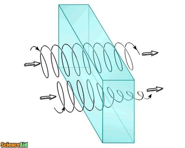
- 1A light which oscillates in a single plane is called linearly polarized light. All polarized lights can be defined as a sum of two linearly polarized lights at right angles to each other, namely the vertically polarized light and the horizontally polarized light. If one polarized light is out of phase with the other by a quarter wave, the resultant will be a circularly polarized light [1].Polarized Light.
- 2A chiral molecule absorbs left circularly polarized light and right circularly polarized light differentially. This difference in absorption is measured by CD spectroscopy. Certain structures have a signature absorption pattern and these known spectra can be utilized to analyze the structure of unknown chiral molecules. (I) The two components have the same amplitude and when combined generate plane polarized radiation (II) the components are of different magnitude and the resultant (dashed line) is elliptically polarized.Chiral molecule.CD occurs only at wavelengths that can be absorbed by the chiral molecule.


Mathematical Representation
In practice, the CD instrument (spectropolarimeter) displays the dichroism at a given wavelength of radiation expressed as either the difference in absorbance of the two components
(DA = AL - AR) or as the ellipticity in degrees
(q) (q = tan-1(b/a), where b and a are the minor and major axes of the resultant ellipse).
The simple numerical relationship between
DA and q (q in degrees) is q = 32.98 DA.
A CD spectrum is obtained when the dichroism is measured as a function of wavelength. In most biological work, observed ellipticities are of the order of 10 millidegrees.
Proteins and Light
Proteins can absorb light in the near UV (320-260nm) and far UV (240-180nm) regions. In far UV CD, the dichroism is due to the chirality of the peptide bond which gives information on common secondary structures like α helix and β-pleated sheets. Near UV signals are weak because only a small number of aromatic amino acids or prosthetic groups contribute to the CD signals in near UV region. The tertiary folding of a polypeptide chain places these aromatic side chains in the chiral environment, giving rise to a CD spectra that is typical for the native structure. The presence of disulfide bonds in proteins can also be detected from signals in the 240-290nm range. Different forms of regular secondary structures in peptides and proteins display characteristics spectra:


Far UV CD spectra associated with various types of secondary structure: solid curve, a-helix; long dashes, antiparallel b-sheet; dots, type I b-turn; dots and short dashes, irregular structure.
- 1Units for CD measurement:
In terms of absorbance, the units are those of the difference in
molar absorbance De = eL - eR , i.e. cm-1.M-1.
In terms of ellipticity, the mean residue ellipticity [θ]mrw, λ at a wavelength λ is quoted in
units of deg.cm2.dmol-1 and is given by:-
[θ]mrw,λ = MRWθ / 10d.c
where q is the observed ellipticity (degrees), d is the pathlength (cm) and c is the concentration (in units of g/ml).
- 2It has a source of linearly polarized monochromatic light which can be converted to left circularly polarized light or right circularly polarized light by passing it through a quarter plate tilted at an angle of 45º to the plane of the linearly polarized light.CD spectropolarimeter.
- 3A circular dichroism spectrophotometer has a specialized optical element called a photo-elastic modulator (PEM); a piezoelectric element cemented to a block of fused silica. When the piezoelectric element oscillates at its resonance frequency (typically around 50 kHz), it induces stress in the silica in such a way that it becomes birefringent (having two different refractive indices). The alternating stress causes the fused silica quarter plate to retard the vertical with respect to horizontal components of the incident linearly polarized light by a quarter-wave to produce left-circularly polarized light(CPL) and then vice versa. The amplitude of the oscillation is adjusted according to the wavelength of light passing through the silica block.A circular dichroism spectrophotometer.
- 4A light detector on the other side of the sample records the light intensity hitting the detector. When a circularly dichroic sample is placed in the light path, the recorded light intensity will be different for right- and left-CPL. The difference in intensity between the two circular polarizations (vAC) is measured by a lock-in amplifier tuned to the frequency of the PEM.A light detector.
Referencing this Article
If you need to reference this article in your work, you can copy-paste the following depending on your required format:
APA (American Psychological Association)
Understanding Circular Dichroism. (2017). In ScienceAid. Retrieved Apr 25, 2024, from https://scienceaid.net/Understand_Circular_Dichroism
MLA (Modern Language Association) "Understanding Circular Dichroism." ScienceAid, scienceaid.net/Understand_Circular_Dichroism Accessed 25 Apr 2024.
Chicago / Turabian ScienceAid.net. "Understanding Circular Dichroism." Accessed Apr 25, 2024. https://scienceaid.net/Understand_Circular_Dichroism.
If you have problems with any of the steps in this article, please ask a question for more help, or post in the comments section below.
Comments
Article Info
Categories : Physics
Recent edits by: SarMal, Sharingknowledge, Adasolutions


