The Brain
Edited by Jamie (ScienceAid Editor), Taylor (ScienceAid Editor)


Cerebral Hemispheres
The cerebral hemispheres are the two main portions of the brain, and make up its majority. An important thing to note is that the left side of the body is controlled by the right cerebral hemisphere, and the right side of the body is controlled by the left cerebral hemisphere.
The different parts of the cerebral hemisphere are sized according to the complexity of their innervation. This means that a more complicated function requires a larger portion of the cerebral hemisphere to perform it. For example, the visual areas take up a large chunk of the back of the brain.
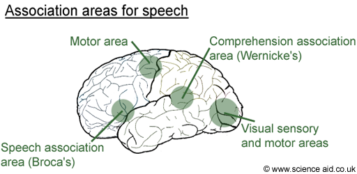
The brain is made up of sensory, association and motor areas.
- 1There are different sensory areas for the different sensory organs.Sensory areas receive and process information from sensory organs.Advertisement
- 2For example, the visual association area interprets what you see, by comparing it to things you've seen before.Association areas interpret the sensory information by relating it to memory.
- 3Motor areas are also localized with certain areas for different movements.
As way of example, the diagram below outlines the different areas that relate to speech.

Autonomic Nervous System
The autonomic nervous system is a branch of the nervous system that deals with things under subconscious control. It has two types of antagonistic (opposite and opposing) nerves:
- 1Sympathetic - which prepares the body for action, such as increasing heartbeat.
- 2Parasympathetic - which does the reverse and returns the body to normal.
In the below table, we look at some reflexes and how they are controlled by the autonomic nervous system.
| Reflex | Action | Explanation |
|---|---|---|
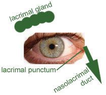
| Pupil diameter | Pupil dilates to allow more light into the eye and constricts to reduce it. | See the eye |
| Tear production |
|
The tear duct sits below the eyebrow. When the sympathetic system is stimulated, tears are produced and secreted by the eye to lubricate and protect it, the film of tears is spread across it by blinking. Extra tears are drained through the nasolacrimal duct to the digestive system. When the parasympathetic system is stimulated, tear secretion is increased. |
| Bladder emptying | The internal and external sphincter muscles open and the detrusor muscle contracts to allow urine out via the urethra. | When the bladder is stretched, impulses are sent by the parasympathetic system to contract the detrusor muscle which surrounds the bladder, and tightens it. In order for the urine to exit; the internal (autonomic control) and external (voluntary control) sphincter muscles must open. Sphincter muscles form a ring, and when they relax, this ring opens. The sympathetic system relaxes the bladder. |
Referencing this Article
If you need to reference this article in your work, you can copy-paste the following depending on your required format:
APA (American Psychological Association)
The Brain. (2017). In ScienceAid. Retrieved Apr 19, 2024, from https://scienceaid.net/biology/humans/brain.html
MLA (Modern Language Association) "The Brain." ScienceAid, scienceaid.net/biology/humans/brain.html Accessed 19 Apr 2024.
Chicago / Turabian ScienceAid.net. "The Brain." Accessed Apr 19, 2024. https://scienceaid.net/biology/humans/brain.html.
If you have problems with any of the steps in this article, please ask a question for more help, or post in the comments section below.
Comments
Article Info
Categories : Humans
Recent edits by: Jamie (ScienceAid Editor)