The Heart and Lungs
Edited by Jamie (ScienceAid Editor), Taylor (ScienceAid Editor), Jen Moreau, Sharingknowledge and 2 others


The Heart

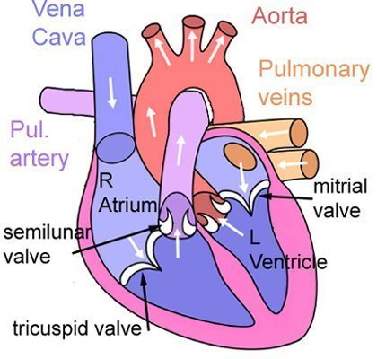
Above is a diagram of the heart. At the top are the entrances or atria. Into these, come the Vena Cava bringing blood from all of the body; and the pulmonary vein bringing blood from the lungs.
Below these are the ventricles, they are separated from the atria by the semilunar valve on the right and the mitral valve, also called the bicuspid or left atrioventricular valve, on the left. The blood is pushed up out of the heart from these.
The Heartbeat
Heart tissue is myogenic which means it will naturally contract and twitch by itself, but the heart needs to coordinate this so that it doesn't just beat and twitch all over the place.

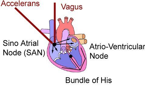
A nervous impulse is sent to the sino-atrial node (SAN) which causes the atria to contract first, then there is a delay where it is received at the atrio-ventricular node which sends the impulse along the Bundle of His to make the ventricles contract, they are said to be in systole. And when they relax they are in diastole.
Now we look at the role of the two nerves connecting to the SAN. These go up to the brain (Medulla Oblongata) and regulate the speed of the heart. The accelerans is like the accelerator in a car, and makes the heart beat faster, and then the vagus tells it to beat slower. In order to get a perfect speed, the signals from either alternate in different amounts. So to get a steady speed half the signals would come from each one.
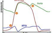
Now we'll have a look at the pressure changes that take place in the heart during the heartbeat. Below is a graph that shows this.

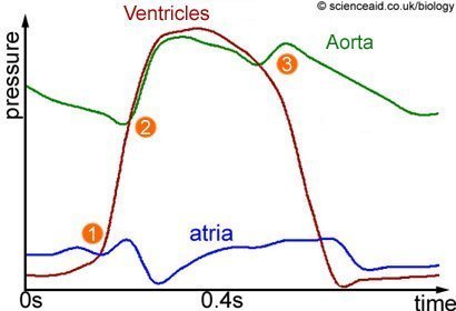
The red line shows pressure in the ventricles. At 1, there is atrial systole (atria contract), this means that blood is suddenly forced into the ventricles; thus increasing the pressure.
Now we look at what happens at 2, the pressure in the aorta represented by green, begin to climb because the semilunar valves have opened since the pressure in the ventricles is enough from ventricular systole. So now blood is being forced into the aorta.
At point 3, both the atria and ventricles are in diastole and relaxing. There is a blip in the pressure of the aorta as the semilunar valve closes and then its pressure falls. The heart relaxes until the next impulse from the SAN starts the whole process all over again.
Questions and Answers
Explain the reasons for regular changes in point 3?
Why is blood pressure high in ventricles? How is the graph helpful?
During Diastole, blood fills the heart and the ventricle then relaxes. This is apparent from the graph (pressure against time). The red line denotes an abrupt change in the ventricular pressure and can be observed when the pressure decreases at point 3, which confirms that the ventricles are relaxing.
Referencing this Article
If you need to reference this article in your work, you can copy-paste the following depending on your required format:
APA (American Psychological Association)
The Heart and Lungs. (2017). In ScienceAid. Retrieved Apr 18, 2024, from https://scienceaid.net/biology/humans/heart.html
MLA (Modern Language Association) "The Heart and Lungs." ScienceAid, scienceaid.net/biology/humans/heart.html Accessed 18 Apr 2024.
Chicago / Turabian ScienceAid.net. "The Heart and Lungs." Accessed Apr 18, 2024. https://scienceaid.net/biology/humans/heart.html.
If you have problems with any of the steps in this article, please ask a question for more help, or post in the comments section below.
Comments
Article Info
Categories : Humans
Recent edits by: Tosin Emmanuel, Sharingknowledge, Jen Moreau


